Thursday, December 8, 2016

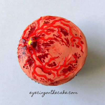
central retinal vein occlusion

What is a CRVO?
A central retinal vein occlusion (CRVO) occurs when there is an obstruction of the central retinal vein, which is the main vein of the retina (the tissue that lines the back of the eye). A blood clot, or thrombus, may occur in the vein as a result of abnormalities in blood vessel size, blood composition and/or blood flow.

Veins carry blood back to the heart. When the main vein that drains the retina is blocked and blood cannot flow out, the blood builds up and leaks out of the walls of the vessels.  This leakage causes the retina to swell (retinal edema).  When fluid leakage occurs in the area of the retina called the macula (macular edema), central vision is impaired.  

We'll be talking specifically about central retinal vein occlusions in this post, but you can also have a branch retinal vein occlusion (BRVO), which affects a smaller vein, or a hemi-retinal vein occlusion (HRVO), which affects either the upper or lower half of the retina. It all just depends on the location of the blood clot. 

A CRVO can be classified as non-ischemic or ischemic.  Ischemic means there is a shortage of oxygen due to a reduced or restricted blood supply.  When ischemia exists in the retina, vascular endothelial growth factors (VEGF) are released. VEGF stimulates the growth of new, abnormal blood vessels (neovascular membranes) to help supply the retina with the necessary oxygen and nutrients.  This is bad, because those blood vessels are weak and leak. In the case of ischemic CRVOs, the abnormal vessels can cause a serious type of glaucoma, called neovascular glaucoma. This results in dangerously high eye pressure that is very difficult to treat. Fortunately, most cases (about 75%) of CRVOs are non-ischemic, which is the less serious form and involves less severe vision loss.  
Photos of a non-ischemic CRVO of the right eye
What are the symptoms and signs?
The most common presentation is sudden, painless loss of vision in one eyeWhen the eye doctor looks in the affected eye, he/she will see lots of hemorrhages (see photos) in all 4 quadrants of the retina and possibly cotton wool spots, which are fluffy white spots in the retina. The veins of the retina are widened and curly. There may also be swelling of the optic disc. CRVOs are sometimes described to have a "blood and thunder" appearance, because it pretty much looks like something exploded in the back of the eye. 
CRVO photo via Wills Eye
What are the risk factors?
CRVOs usually occur in persons over the age of 50, and hypertension is the most common systemic association. Other vascular diseases like atherosclerosis, high cholesterol, and diabetes are risk factors as well.  Hardening and/or narrowing of the arteries can compress the vein and cause clot formation.  Open angle glaucoma is also a risk factor.  If a CRVO occurs in someone under 40 years of age with no known risk factors, he/she may need to be tested for blood clotting or thickness abnormalities

How is it treated?

The blockage cannot be undone, so the goal is to treat/prevent the secondary complications. Namely, macular edema and neovascularization. Several clinical trials have shown there are treatment options that help reduce macular edema and improve vision to a certain degree. Early detection of complications and timely treatment are key.  

The most common treatment for macular edema as a result of a CRVO is intravitreal injections of anti-VEGF drugs. The drugs are injected into the gel part of the inner eye (the vitreous), and are usually administered every 4-6 weeks.  Two such drugs that are FDA-approved for treating macular edema due to CRVOs are Lucentis (ranibizumab) and Eylea (aflibercept). Avastin (bevacizumab), a cancer drug, is an anti-VEGF drug used extensively as well, though used off-label. Steroid injections/implants may be used instead of or in addition to anti-VEGF injections. An intravitreal steroid implant, Ozurdex, is an FDA-approved treatment for macular edema secondary to vein occlusion. Currently in FDA trials: a combo of Eyelea (intravitreal anti-VEGF injection) and Zuprata (suprachoroidal steroid injection) that has the potential to decrease the number of Eyelea injections a patient needs (read more about it here). Laser is not a common treatment with CRVOs, unless there is neovascularization present.

Macula OCT of the above-photographed CRVO patient
Aside from taking photos to help monitor the condition, your eye doctor may utilize other tools in the treatment process. 

  • Optical coherence tomography (OCT) is a scanning laser used to get a cross-sectional image of the retina (see scan above).  This helps assess the level of swelling in the macula and track the response to treatment.  
  • Fluorescein angiography (FA) is used to identify areas of the retina with poor/absent blood flow and helps guide treatment. A dye is injected into a vein in the arm and photos of the retina are taken as the dye reaches the retinal vessels.


CliffsNotes:  A CRVO occurs when a blood clot blocks the outflow of blood from the tissue that lines the back of the eye (the retina). If you notice a sudden, painless loss of vision, contact your eye doctor STAT!


Wednesday, October 19, 2016

Demodex

What is Demodex?
Demodex is an 8-legged, microscopic mite. Two species live on humans: Demodex folliculorum, which are found in the eyelash follicles, and Demodex brevis, which are found in the sebaceous (sweat) and meibomian (oil) glands. These mites live primarily on the face and feed on skin cells and oil in the follicles and glands. They avoid sunlight and live for about 2 to 3 weeks. How common are they? Fairly common in adults (studies report they are found on the skin of 20-80% of adults- that's a pretty wide range and not a super helpful stat...), and nearly 100% of elderly people have the mites on their skin (1). Studies have shown that the prevalence of Demodex increases with age, and Demodex mites are present in higher numbers in those with rosacea, compromised immune systems, and possibly contact lens wearers (23).


Two D. folliculorum on a plucked eyelash (A) and one hanging solo (B)
image: JKMS

Fun (or disgusting) fact: Demodex mites have no anus. Their waste is stored in the gut and expelled when they die, which may cause additional inflammatory response. Shout-out to one of my patients who shared that fact with me. #mypatientsaresmarterthanme

Demodex being teased out of the follicle (arrow) via lash rotation
image: Optometry and Vision Science

What problems can Demodex cause?
Many people have Demodex mites on their skin and have no symptoms or complications. However, Demodex overpopulation (demodicidosis) has been linked to (not necessarily causative of) skin conditions such as blepharitis, rosacea and perioral dermatitis, as well as meibomian gland dysfunction, dry eye, recurrent chalazion, and ocular surface inflammation (45).

Demodex mites can cause problems by clogging hair follicles and sweat ducts, causing abnormal thickening and growth of the epithelial cells at the lid margin. Further, the exoskeleton and waste of the mites can cause an inflammatory response (67). Overpopulation of Demodex mites has been linked to an increase in the presence of certain cell-signaling proteins (cytokines, specifically interleukins) in the tears, which can cause an inflammatory or immunological response (8).

The presence of cylindrical dandruff or "waxy sleeves" around the base of the eyelashes is a very good indicator of demodicidosis. Symptoms may include itching and redness of the lid margin, loss of lashes or misdirected lashes, as well as burning or irritation.

Cylindrical flakes that are highly suggestive of demodex
image: Review of Optometry

How do you get rid of the little buggers?
Frequent scrubbing of the lids with diluted tea tree oil (TTO) was found to be the most effective way to eradicate Demodex and improve patient symptoms (9). The TTO has ingredients (namely, terpinen-4-ol) that kill mites and ticks while also luring the mites out of the hair follicle before they mate (10). There are also lid wipes, scrubs and shampoos containing various concentrations of TTO that can be used for at-home treatment and long-term maintenance. It is best to keep the concentration of TTO at or below 50%; any higher than that may be too irritating (11). Your eye doctor may choose to add an antibiotic and/or steroid ointment or drop to the treatment as indicated, depending on what other symptoms and signs are present.


CliffsNotes: If you suffer from dry, itchy, irritated, red, and/or crusty eyelids, have your optometrist take a look. You MITE (see what I did there?) have a Demodex party going on in your eyelashes. 


Additional Recommended Resources:

Thursday, September 22, 2016

the contact lens case

As eye care professionals, we talk a lot about the proper care of contact lenses, but we probably don't spend enough time talking about the proper care of the contact lens CASE. Sure, it doesn't actually go ON your eyeball, but it does matter! The contact lens case can act as a reservoir for microorganisms and can be the source of eye infections, with the contact lens serving as the vector.  Some microbes can attach to the case and secrete substances that create an extracellular matrix-type protective encasement called a biofilm. This allows adherence to biological tissue, plastics, medical devices, etc. The ideal environment for biofilms to attach and grow is a moist surface, which makes contact lens cases prime real estate (1). 

So how should you be taking care of your contact lens case?

1. Change the case AT LEAST every 3 months.  Look at the photos below. Crystal violet stains protein and DNA, the primary components of cellular debris.  You can see that the intensity of the staining correlates with the age of the lens case (2). The longer you use the case, the more bacteria and debris collects, which increases your risk of infection.


image: RCCL

2. Clean the case daily by rubbing and rinsing with fresh contact lens solution (not water). A study found that 52% of those questioned cleaned their cases with tap water (3).  Why is that bad?  Because tap water contains microorganisms that can lead to rare but serious eye infections, like Acanthamoeba keratitis. In another study, cleaning cases with tap water was found to be associated with a higher rate of contamination with gram negative bacteria (4). Multipurpose solutions are designed to interfere with the cell membrane of microorganisms and kill them. Hydrogen peroxide-based solutions use a different method to disrupt the microbial membrane, as well as the DNA and other cellular components (5).  

3. Wipe the contact lens case with a clean tissue, and place the case upside down to air dry in a clean area (ie: not inches from the toilet). Simply rinsing with contact lens solution is not enough to destroy biofilms; mechanical disruption is needed (37).  More bacterial contamination was found to occur in humid environments, most notably when the case was left to air dry face up (8).  So the research tells us that the most effective way to clean your contact lens case is to rub and rinse the contact lens case with fresh contact lens solution daily, wipe the case with a clean tissue, and leave it face down on a clean surface to air dry (910).  

The proper way to store your case: upside down on a clean tissue.

4. Be sure to keep the contact lenses themselves clean!  More on that in a previous post.

Even better yet? Get rid of the case all together and opt for daily disposable contact lenses. You put them in in the morning, remove and discard them in the evening, and start with a fresh pair the next day. Talk to your optometrist to see if those are an option for you.

CliffsNotes: Rub and rinse the contact lens case with fresh contact lens solution (not water) daily, dry with a clean tissue, and leave upside down on a clean tissue to air dry. RUB, RINSE, TISSUE-DRY, AIR-DRY. Replace the case at least every 3 months. 

Additional Resources:


Thursday, September 1, 2016

the history of eyeglasses

A few weeks ago, I saw a 16 year old patient that was interested in optometry as a potential career path. He asked a few questions about the field and schooling, then he asked me a question that stumped me: "When were glasses invented?"

::Crickets:: I am admittedly not a history buff, but this was one I probably should have known. So this blog post is for you, inquisitive teenager S.

When were eyeglasses invented?
Short answer: In the 1200s.

Long answer: The invention of the eyeglass is thought to have occurred in Italy between 1268 and 1289. The use of glass as a means of enhancing vision was mentioned in a few manuscripts around this time. One such mention was in a book by Robert Grosseteste printed in 1235. It referenced using a glass to “read the smallest letters at incredible distances.” In 1268, the English philosopher Roger Bacon wrote: "If anyone examine letters or other minute objects through the medium of crystal or glass or other transparent substance, if it be shaped like the lesser segment of a sphere, with the convex side toward the eye, he will see the letters far better and they will seem larger to him. For this reason such an instrument is useful to all persons and to those with weak eyes for they can see any letter, however small, if magnified enough."

In 1289, Sandra di Popozo wrote in a manuscript: "I am so debilitated by age that without the glasses known as spectacles, I would no longer be able to read or write. These have recently been invented for the benefit of poor old people whose sight has become weak." In 1306, a monk of Pisa delivered a sermon in which he stated: "It is not yet twenty years since the art of making spectacles, one of the most useful arts on earth, was discovered. I, myself, have seen and conversed with the man who made them first." Thus, we conclude that eyeglasses arrived on the scene in the mid-1200s.

The first artistic rendering of spectacles was found in a fresco of Cardinal Hugo of Provence painted by Tommaso da Modena around 1352.
Tomasso da Modena's painting
image: History of Information

In their early days, glasses were worn primarily by monks and scholars. They were essentially two pieces of glass or crystal riveted together, with the lenses bound in leather, metal, or bone. They were either held in front of the eye or balanced on the nose. It wasn't until the invention of the printing press around 1440-1450 that eyeglasses became a common item.

Early spectacles, framed in leather
image:AAO Museum of Vision

The idea of attaching ribbons of silk to the frames and looping them over the ears emerged in the 17th century by Spanish spectacle makers. This idea made it over to China by way of Spanish and Italian missionaries. The Chinese modified this, adding little ceramic or metal weights to the string to hold it in place behind the ear, instead of using a loop. Edward Scarlett, an optician in London, is credited with creating the rigid sidepiece that rested over the ears in 1730. Thus, the spectacle temple was born.
Edward Scarlett's trade card, the first advertisement of rigid temples 
image: The College of Optometrist Museyeum
Martin's Margins 
image: AAO Museum of Vision
Binocles-ciseaux, or scissor-glasses, reached the height of their popularity in the latter half of the 18th century. George Washington and Napoleon Boneparte are said to have used these.
Scissor-glasses 
image: AAO Museum of Vision
Another form of spectacles that came about in the 18th century was the lorgnette, a pair of lenses that were held in front of the eyes by a side handle. These may have developed from the scissor-glasses. Created by George Adams, lorgnettes often had ornate and embellished handles, as they were worn mostly by fashionable women.
The lorgnette (everyone should have a fan attached to their specs) 
image: AAO Museum of Vision
The monocle, interestingly called an "eye ring," become popular in the mid to late 1800s, though they are thought to have developed in Germany earlier. The monocle was most often worn by upper class men. And, of course, Mr. Peanut.
The monocle- who wore it best?
image:Wikipedia
The 1800s saw the oval frame shape come to prominence, as well as the emergence of bifocals. Benjamin Franklin is widely credited for the invention of the bifocal in the 1780s, though the jury is still out on that one.  B.M. Hanna (Hannas unite!) received patents for the cemented and perfection bifocals in the late 1800s.

Pince-nez is a style of glasses that came about in the 1840s. French for "pinch nose," these specs literally pinched the nose and usually had a chain that attached to a lapel or dress. President Teddy Roosevelt was often pictured wearing these. (Aside: check out this interesting story about how President Roosevelt's eyeglass case helped him dodge a bullet.)

Speak softly and carry a big stick. And wear your pince-nez.  
image:Wikipedia
Another interesting tidbit for my eye nerds out there: J. J. Bausch emigrated to the US from Germany in 1850. He had apprenticed with an optician and came to America in search of better opportunity. He set up shop in NY as an optician, and struggled for many years. He sought a $60 loan from his friend, Henry Lomb, and Lomb became an apprentice under Bausch. Bausch created frames out of vulcanized rubber, which were stronger and less expensive than the current metal, animal horn, or wood frames out there. Demand for the product increased, and J.J. Bausch and Henry Lomb eventually went on to build the Bausch and Lomb Optical Company. For those of you that may not know, Bausch and Lomb still exists today, making it one of the oldest continuously operated companies in the US (more info here and here).

The 1900s saw eyeglasses become stylish in addition to functional. A wider variety of eyeglasses were available, and the stigma around wearing eyeglasses was beginning to fade away. Movie stars became style icons, and their glasses were part of their image. Sunglasses also became popular in the 30s. Progressive addition lenses (PALs) were invented in the 50s.
Comedian Harold Lloyd, "the man who popularized eyeglasses in America," wearing his iconic horn-rimmed glasses.
image: Wikipedia

And here we are, in the present, where eyeglasses are considered part of a person's wardrobe, an accessory even. Some old trends are coming back, and some new ones are being created. There are now more eyeglass styles and colors than you can dream of, and they are such a fun (and useful) form of self-expression.

CliffsNotes: Eyeglasses were invented in Italy in the 1200s. They've come a long way since then. :)

Sources
:

Thursday, July 14, 2016

4 reasons NOT to buy glasses online

four-eyed cupcakes

Every now and then, I have patients tell me that they plan on buying their glasses online.  I can certainly understand why that would be appealing: low prices, large selection, and ease.  In our drone-delivery, instant-gratification culture, convenience is king!  I, personally, am all about finding cheaper prices from the comfort of my Snuggie (before you start judging, it's a Buzz Lightyear Snuggie).  However, knowing what I know about vision and eyewear, I would urge you to think twice before buying glasses online.  In pursuit of convenience and ease, we forgo the knowledge and expertise of the eyecare professional team in precisely fitting and knowledgeably selecting our eyewear.  This blog post is my attempt to "let the buyer beware."

----------

Here are 4 things to consider when looking into buying eyeglasses online:

1. Questionable lens accuracy and quality. For me, this is the biggest reason to avoid buying glasses online.  A recent study of internet eyewear orders found that nearly half (44.8%) of the glasses examined had incorrect prescriptions or safety issues:
  • Nearly three out of 10 pairs (29%) of glasses ordered online had at least one lens that failed to meet the required prescription.  When you buy a pair of glasses from an optometrist's office or optical, someone typically checks the lenses for accuracy and quality before the glasses are dispensed to you.
  • Nearly a quarter (23%) of the lenses failed impact resistance testing, which highlights a major safety issue.  Children’s glasses performed even worse, with 29% failing impact testing. (1)
Quality and safety are a big deal, as is prescription accuracy.  These are not areas where you want to cut corners.  At a very minimum, you need to be sure that the power in your lenses is the power on your prescription.  I have had more than a few patients come in complaining about the glasses they bought online, only to find that the prescription in their lenses is in fact not the prescription I wrote for them.  And there's more to it than just having the prescription correct; you can still have headaches, eyestrain, and problems seeing even if the numbers match.

Beyond having the correct power in your lenses, you also need to be looking through the right part of the lens to see clearly and comfortably.  The optical center of your lens should be placed right in front of the center of your pupil.  For the maker of the glasses to know where exactly to put the lens within the frame to achieve that, an accurate pupillary distance is needed.  Multi-focal lenses (lenses that allow you to see at more than one distance, like bifocals or progressive addition lenses) involve additional measurements and powers; fitting multifocal lenses is a careful process and should definitely be done by experienced professionals.  The segment height is a specific measurement for multifocal lenses, and it needs to be measured while you are wearing the frame, with the frame positioned where you normally wear it.  Both the pupillary distance and the segment height are very important measurements that influence what part of the lens you are looking through, and what power you are getting when looking through that part of the lens.  The more complicated the prescription, the more crucial it is to consult with experienced professionals.  Some forms of optical correction, especially for children, are prescribed as part of a treatment for a condition, such as accomodative esotropia or accomodative insufficiency.  If given the incorrect treatment, the condition will not be effectively treated.  After all, glasses are considered Class 1 Medical Devices by the Food and Drug Administration (FDA).  Glasses are not just an accessory; they require a precise prescription and accurate measurements to enable you to see clearly and comfortably.

2. Lack of customization.  Health care should be pursued in-person, because it should be tailored to the individual.  What you need in eyewear depends on your specific prescription and visual needs.  A conversation with your optometrist and optician about such things should happen in order to build a pair of glasses that gives you the best vision and comfort for your daily life.  Do you use a computer all day?  Do you drive for a living?  Do you knit?  Do you play golf?  Now more than ever, there are SO many choices in terms of lens types, materials, and coatings; it really is important to speak with a knowledgeable expert to help you navigate the options and decide on what products suit your visual needs and increase your visual comfort.

3. Lack of input in selecting an appropriate frame.  Frame selection is both an art and a science.  Did you know frames have sizes?  Glasses are NOT one-size-fits-all.  If glasses don't fit properly, you can experience physical as well as visual discomfort.  Aesthetically speaking, certain frame shapes look better on certain face shapes.  But more importantly, there are some frame sizes and styles that should be ruled out based on your prescription and/or the type of lens you need. A trained optician can tell you what frames to avoid.  The way a frame looks certainly matters, but there are many other factors to consider when choosing a frame for your lenses.

4. Customer service.  This is by no means a rule, but in general, the smaller the shop, the better the customer service. There are typically only a few degrees of separation, if any, between you and the manager or owner. You have a person to go back to if you are having any issues with your glasses, or if you need a frame adjustment. I believe human interaction is SO important to building business relationships and creating loyalty. Maybe I'm an old soul, but it's just not the same as clicking around on a website or calling a 1-800 number.

----------

Every pair of glasses purchased online is not a disaster.  But I would suggest that you as a consumer consider service and quality as well as price and convenience.  Glasses are an investment, and you want your money to go towards a pair that fits properly and comfortably, and provides you with the best possible vision. If you choose to purchase your eyeglasses online, be informed and may the odds be ever in your favor! :)


CliffsNotes: Glasses are not one-size-fits-all accessories. A team of eye care professionals can help you choose a frame and lenses that are ideal for you, considering your prescription and visual needs.  In my opinion, the convenience of ordering glasses online is not worth the potential compromise in accuracy and quality.


Additional recommended resources:

Thursday, May 19, 2016

cancer and the eye

I would venture a guess that everyone reading this blog has been affected by cancer, either personally suffering from it or having a loved one that is suffering/has suffered from it. That makes it a pretty relevant topic to discuss here. Eye cancer is rare, certainly, but it does exist.  Most people don't see the eye as an organ, just like the lungs or liver, that can have tumors.  In terms of eye tumors, there are many types of benign (harmless, non-invasive) and malignant (bad news bears) tumors that can be found in any part of the eyeball and surrounding tissue.  For the sake of brevity, we won't talk too much about the benign tumors; instead, this post will just focus on the scary stuff.

First off: what is cancer?  Cancer is a group of diseases characterized by uncontrolled cell division. These dividing cells may form tumors.  Malignant tumors are cancerous; they can invade nearby tissue. Cancer originates in one place and can spread to other parts of the body. The term for this spread is metastasis.  The National Cancer Institute has a great, easy-to-understand overview of cancer here. According to the Center for Disease Control (CDC) and the American Cancer Society (ACS), the most common cancers in the US are breast, prostate, and lung (1, 2).

From ACS

In terms of ocular cancer, there are various sites within and around the eye that cancerous tumors can be found. We'll go through some of the major types in each category.

1. INTRAOCULAR [within the eye]
  • Primary intraocular cancers are cancers that originate in the eyeball.
    • Retinoblastoma is the most common eye cancer in children, accounting for 3% of all childhood cancers. This rare cancer can be non-inherited or inherited via a mutation in the RB1 gene, which is also linked to an increased risk of osteosarcoma (bone cancer). All babies are screened at birth, but this may not be present at birth. That's why it is crucial to not only see your pediatrician for well-baby exams but to ALSO see your eye doctor (for more info on infant eye exams, read the InfantSEE post). Early diagnosis is key!  The most common signs of retinoblastoma are a white pupillary reflex (instead of the normal red reflex that you often see when you take a flash photo) called leukocoria, and eye turns (strabismus) (3)
Leukocoria from retinoblastoma, image: University of Michigan
    • Uveal melanoma is the most common primary intraocular malignancy in adults. It forms in the eye's melanocytes, the cells that make pigment (melanin). [The uvea is the pigmented middle layer of the eye and includes the iris, ciliary body, and choroid.  The choroid is the vascular tissue that lies between the retina (the light sensitive tissue that lines the back of the eye) and the sclera (the white part of the eye).] Choroidal melanomas account for 80% of all uveal melanomas, affecting six out of every one million Americans (4). Choroidal melanomas most often have no symptoms and are found during routine eye examination. Choroidal melanomas can metastasize, to the liver most commonly, also occasionally to the lungs, bone, skin, and brain (Kanski, 7th ed)
    Choroidal melanoma, image: ECN
    • Primary intraocular lymphoma (PIOL) often presents in the elderly and immunocompromised, with possible symptoms of floaters, blurred vision, redness, and/or sensitivity to light. Lymphoma is a cancer of the lymphatic system, the system that helps protect the body from infection. Lymphoma is classified as either Hodgkins or non-Hodgkins.  Non-Hodgkins lymphoma can be further categorized based on what type of cells are involved.  Most are B-cell.  Because lymph tissue is found throughout the body, lymphoma can begin nearly anywhere. Lymphomas can occur intraocularly but also can occur in other structures on/around the eye (more on that later).  PIOL is primarily non-Hodgkins, B cell lymphoma, and is highly correlated with primary central nervous system lymphoma (PCNSL). In fact, up to 80% of patients with PIOL will be subsequently diagnosed with brain lymphoma (5).
  • Secondary intraocular, or metastatic, cancer is cancer that originates elsewhere in the body and spreads to the eye.  Secondary intraocular cancer occurs more often than primary.  The most common cancers to spread to the eye are breast and lung, and the most common place for them to spread within the eye is the vascular choroid.  One study found that 34% of those eyes diagnosed with uveal metastasis did not have a history of cancer at the time of diagnosis. Further evaluation of that 34% revealed over a third of them had a primary tumor in the lungs (6).  According to Wills Eye Hospital, about 25% of patients who present to the eye doctor with eye metastasis have no known history of systemic cancer and are later found to have a cancer in the body (7).  Key point: the detection of cancer in the eye can precede the diagnosis of cancer elsewhere in the body, especially in the case of lung cancer. 

2. CONJUNCTIVA [the tissue covering the white part of the eye]
  • Conjunctival squamous carcinoma occurs most often in older white males, and is linked to UV exposure. This rarely spreads to distant sites but can spread locally behind the eye and in the orbit (8).
  • Conjunctival melanoma appears as a pigmented area on the white part of the eye. It can arise from an existing nevus/mole or from a precancerous condition called primary acquired melanosis (PAM).  Unlike a squamous carcinoma, there is a risk of spread to the lymph nodes and other organs (9)
  • Conjunctival lymphoma presents as a salmon-colored patch, often found under the eyelids, most commonly in late adulthood. In about 20% of cases, lymphomas in other parts of the body are found (10).
Conjunctival lymphoma, image: Wills Eye

3. EYELIDS [The Skin Cancer Foundation reports that eyelid skin cancer accounts for 5-10% of all cases of skin cancer.]
  • Basal cell carcinoma (BCC) is by far the most common eyelid tumor, accounting for about 90% of eyelid cancers. Most occur on the lower lid, and are found more commonly in older, fair-skinned people with chronic exposure to UV radiation. BCC is locally invasive but does not metastasize (Kanski, 7th ed).
  • BCC, image: Wills Eye
  • Squamous cell carcinoma (SCC) is similar to BCC in that it's typically found on the lower lid of older, fair-skinned patients with chronic exposure to UV radiation.  It is less common than BCC, but more aggressive, and metastasis to nearby lymph nodes occurs in 20% of cases (Kanski, 7th ed).
  • Melanoma accounts for less than 1% of eyelid cancers.  Melanomas are aggressive and potentially life-threatening. They are typically pigmented, but sometimes not.  Melanomas can look like moles (nevi), or can arise from moles. The ABCs of melanomas point out characteristics of malignant melanomas that are usually not present with benign moles:
    • Asymmetry
    • Border (uneven or irregular)
    • Color (non-uniform, variety of colors or shades)
    • Diameter (bigger is not better)
    • Evolution (change over time)
  • Sebaceous gland carcinoma (SGC) is a malignant cancer arising from the oil glands in the eyelids. It typically presents as a painless bump on the upper lid, and can be difficult to diagnose. Sebaceous carcinoma is found more frequently in females than males, and is more common in the elderly (Kanski, 7th ed). It can metastasize to lymph nodes and other organs (11).
  • Merkel cell carcinoma (MCC) is a rare form of skin cancer that grows quickly and metastasizes early. It appears as a raised, red-violet bump on the skin, typically linked to chronic sun exposure. MCC is most common in elderly caucasians (12).

  • MCC, image: pubmed

4. ORBIT [the eye socket, including bones, muscles, nerves, and the lacrimal glands (the almond-shaped glands that secrete tears)]
  • Rhabdomyosarcoma is a malignant tumor that arises from skeletal muscle.  About 10% of cases involve the orbit, usually causing the eyes to bulge (proptosis) (1314).
  • Lacrimal gland lymphoma accounts for over a third of all malignancies of the lacrimal gland, predominantly affecting elderly women (15). It was found that one to two-thirds of patients develop systemic disease (if not already present at the time of orbital biopsy), usually within 2.5 years of orbital biopsy (16).
  • Adenoid cystic carcinoma of the lacrimal gland is the most common malignant epithelial tumor of the lacrimal gland. Occurring most often between the ages of 40 and 60, adenoid cystic carcinoma causes the eye to be displaced downward, and can also cause pain as it spreads to the nearby nerves and bone (17,18).
CT of an adenoid cystic carcinoma of the lacrimal gland, image: ECN
  • Malignant mixed tumor of the lacrimal gland is the second most common malignant tumor of the lacrimal gland. It occurs most often in the elderly. It can appear without a history of lacrimal mass, or it can arise from a prior benign mass that was incompletely excised or a long-standing mass that suddenly increases in size (19. 20).

5. BRAIN
Why do I include this in our conversation?  Because the eyes are an extension of the brain, and some brain tumors can be detected during an eye examination.  I plan to do a separate blog post about it, but in short, visual field exams can help detect and even localize some masses in the brain.


CliffsNotes: Cancer can start in and around the eye, or it can spread to the eye from elsewhere in the body. It often doesn't have symptoms, so be sure to get your yearly dilated eye exam whether you have a history of cancer or not!

Additional recommended resources:

Saturday, April 16, 2016

eye floaters

Ever wonder what those squiggly things are that float around in your field of vision?  They may look like bugs, strands, or cobwebs, and they are especially noticeable when you're looking at a plain, bright background (like a clear sky). When you move your eye to get a closer look, those sly boogers drift away! Wonder no more: they are vitreous floaters.

Let's do a quick anatomy review before we continue: The vitreous (sometimes called vitreous humor or vitreous body) is the transparent, gel-like substance that fills the area between the lens and the light-sensitive tissue that lines the back of the eye (retina).  The vitreous accounts for about 80% of the eye's volume.  It is made up of water (~99% of its volume is water) and a network of proteins (collagen fibrils) and sugars (hyaluronan).  As we age, the vitreous becomes less gel-like and more liquid.  The vitreous is attached to the retina at a few key points.

From Mayo Clinic
What is a floater?
What we see as floaters are actually the shadows cast on the retina by substances in the vitreous.  Those substances can be clumps of protein, blood cells, or pieces of tissue.  Check out this great video for an illustration.

Below are a couple of Optomap images that show floaters (both photos are from the same patient- we decided she just has very photogenic floaters).  Depth doesn't translate well in these photos, so here's an explanation of what we are looking at: if you cut the eyeball in half, the red tissue you see in the photos is the inside lining of the back of the eye (retina), and the strands/blobs that the arrows are pointing to are the floaters in the vitreous, which is in front of the retina.


What causes floaters?
Sometimes, floaters are no big deal.  The floaters that we see from time to time are usually just that; they're clumps of fibers that cast shadows on the retina.  Other times, floaters are a symptom of an eye condition.
  • Posterior vitreous detachment (PVD): A PVD can occur as a result of trauma, though it's most often a result of age-related changes in the vitreous. As the vitreous shrinks and becomes more liquid, it may collapse and pull away from the retina.  The point where the vitreous was attached to the optic nerve is what most people complain of- a large floater that appears somewhat suddenly.  A PVD typically occurs earlier in people who are near-sighted and those who have had cataract surgery or eye trauma (1).  Another symptom that may be experienced during a PVD is flashes of light. The mechanical pulling of the vitreous on the retina as it detaches can cause stimulation of the retinal photoreceptors, resulting in the perception of flashes of light.  The concern with a PVD is that the pulling of the vitreous from the retina may result in a retinal break. Between 8% and 26% of acute, symptomatic PVDs are associated with a retinal tear upon initial examination.  Even if no break is found upon initial examination, there is still a 2-5% chance of a retinal break being found a few weeks later (234). Take home point: it's impossible to know if there is cause for concern based on symptoms alone; a thorough examination is necessary. 
  • Retinal break: A retinal break can occur as a result of a PVD or trauma.  Most retinal breaks are treated, as a break can allow fluid under the retina and cause a retinal detachment, which results in permanent vision loss if untreated
  • Vitreous hemorrhage: The vitreous contains no blood vessels, so blood in the vitreous comes from the leakage of vessels into/near the vitreous. This finding is usually associated with trauma, a PVD (with or without retinal tear, but more often with), or vascular disease. The source of the blood can be breakage of a normal retinal blood vessel, as a result of a tear or trauma, or it can be breakage of an abnormal blood vessel (neovascular membrane).  For example, I had a young patient with insulin-dependent diabetes come in complaining of a cobweb in her vision.  The cause of her symptoms was a large hemorrhage in the vitreous stemming from diabetic retinopathy. She had a neovascular membrane form in the back of the eye; these vessels are inherently fragile and bleed easily.   
  • Vitritis: A vitritis is an inflammation of the vitreous body.  The inflammatory cells in the vitreous can cause floater-like symptoms. Blurred vision is also a common accompanying symptom. A vitritis can occur as a result of an infection, autoimmune disorder, or trauma, or it can be idiopathic (no known cause). This entity requires treatment.

I have floaters! What should I do?
As mentioned earlier, a few floaters every now and then is not unusual.  However, you should see your eye doctor right away if you experience:
  • sudden floaters and/or flashes of light
  • an increase in the frequency and/or number of your floaters 
  • a loss of peripheral vision

How are floaters treated?
Really, they're not.  The underlying cause of the floaters may need treatment, as in the case of a retinal tear, a vitreous hemorrhage, and a vitritis.  PVDs, in the absence of a retinal tear, are just monitored.  Floaters often become less noticeable as they break apart and settle towards the bottom of the eye, and as the brain learns to ignore them. Rarely, there are instances where laser treatment or vitrectomy (a surgery where the vitreous is removed) may be pursued to remove large, very symptomatic floaters. However, risks-vs-benefits must be carefully considered, as floaters are harmless and there are risks associated with any surgical procedures.


CliffsNotes: Occasional floaters are normal and increase with age.  However, if you have sudden floaters and/or flashes of light, a loss of peripheral vision, or an increase in the frequency and/or number of your floaters, it's a good idea to go see your optometrist ASAP.


Additional recommended resources:

Monday, February 22, 2016

age-related macular degeneration

Fundus cupcake sprinkled with drusen
February is Age-related Macular Degeneration (AMD) Awareness Month. AMD is one of the leading causes of irreversible vision loss among Americans over 60 (1)

Let's preface our AMD discussion with a quick anatomy review. The tissue that lines the back of the eye is called the retina. The retina has millions of light-sensing cells called photoreceptors (rods and cones). These cells absorb light and convert it into signals that are sent to the brain via the optic nerve, allowing us to see the world around us. The macula is a small area near the center of the retina that is responsible for our central, sharpest vision. Within the macula is a high density of cones, the photoreceptors responsible for color vision. When you look at retinal photos (or the cupcake above), the macula is the dark red area. For reference, here is a labeled photo of a healthy retina.

What is age-related macular degeneration?
Age-related macular degeneration, commonly called AMD or ARMD, involves a breakdown of the tissue that makes up the macula. Waste products called drusen accumulate beneath the retina, in and around the macula. Drusen are the yellow-ish deposits you see in the photo below. This disruption causes the photoreceptor cells to die, which impairs vision. Over time, central vision becomes blurry and distorted. AMD can be classified as either dry or wet.  
  • Dry (non-exudative) AMD: This is the most common form of the disease. The early and intermediate stages of dry AMD involve drusen and/or pigmentary changes in the retinal pigment epithelium (RPE). The late stage involves the death of large areas of retinal tissue, called geographic atrophy, which affects central vision significantly.
Fundus photo of dry AMD
  • Wet (exudative) AMD: About 10% of dry AMD cases progress to the wet form, meaning that neovascularization has occurred. Neovascularization is the formation of new, abnormal blood vessels under and into the retina. These vessels are weak and can leak, causing vision loss. Neovascularization is bad news bears and warrants prompt treatment (discussed below). Wet AMD typically affects vision more rapidly and more significantly than dry AMD.

What are the risk factors?
AMD is a multifactorial disease. The exact cause is unknown, but research suggests some factors increase the risk of AMD:

  • Age. Age is a major risk factor, with most cases typically occurring after the age of 50. 
  • Smoking. Several studies have found a positive association between smoking and the development of AMD. The risk of developing the disease for current smokers is at least two to three times greater than the risk for non-smokers (2, 3). Smoking is the leading modifiable risk factor for AMD.
  • Family history/genetics. Having a close relative with AMD increases the risk of developing the disease (4). Researchers have identified variants of a few complement genes that are associated with an increased risk of developing AMD (5).
  • Diet. Those with diets low in omega-3 fatty acids and antioxidants may be at greater risk of progression to advanced disease (6).
  • Hypertension. Some studies have shown wet AMD to be associated with moderate to severe hypertension (78).
  • Obesity. Research has suggested an association between body mass index (BMI) outside the normal range and early AMD (9)  as well as progression to advanced AMD (10).
  • High cholesterol. Elevated high-density lipoprotein (HDL) levels may be associated with higher risk of AMD (11).
  • Sun exposure.  Exposure to large amounts of visible and blue light may play a factor in disease progression (121314)
  • Ethnicity. AMD is more prevalent in Caucasians. 
  • Gender. AMD is more prevalent in females.

How is AMD diagnosed?
AMD is diagnosed during a dilated eye exam. It may manifest itself in changes in central vision, but it most likely will not in the early stages. Upon dilation, pigmentary changes and/or drusen may be noted by your eye doctor in/around the macula. 

How it is monitored and treated?
  • Various tools exist to help monitor AMD and guide treatment: 
    • Retinal photo. Periodic photos allow for monitoring progression over time. 
    • Amsler grid. This is an easy way for patients to monitor for changes in their central vision. AMD can cause parts of the grid to appear distorted or missing. I suggest that my AMD patients leave it on their refrigerator and test each eye a few times a week, monitoring for any changes. 
      • The ForeseeHome monitoring device utilizes a similar concept. Patients are presented with a series of straight lines containing a wave/bump, and the patient must identify where the distortion is. The device collects the data and alerts the patient's eye doctor if there are abnormal results. 
    • Optical Coherence Tomography (OCT).  A scanning laser is used to image the tissue of the back of the eye. It can produce a cross section of the macula, showing retinal thinning or thickening.  
OCT showing drusen (red arrows) below the retina

    • Fluorescein angiography (FA). Fluorescein dye is injected into a vein in the arm and photos of the retina are taken as the dye reaches the retinal vessels. An FA may be ordered to identify neovascular membranes, evaluate leakage, and/or guide treatment. 
    • Coming down the pike: OCT Angiography. This technology just got FDA approval in the US. It will allow doctors to image the vessels of the retina and choroid without injecting a contrast dye (unlike an FA). 
  • Treatment: 
    • Dry- There is no FDA-approved treatment for dry AMD as of yet. Plenty of research is being done in this area. Studies have shown that antioxidant supplementation may help slow progression to advanced AMD (1516). For more information on ocular nutrition, check out this post.  
    • Wet- Most cases of wet AMD are treated with anti-vascular endothelial growth factor (VEGF) drugs. Anti-VEGF drugs are periodically injected into the eye to stop abnormal vessel growth. Photodynamic therapy (PDT) and laser photocoagulation are also treatment options, though less commonly used.
Vision loss is often life-altering and difficult to cope with. There are support groups and low-vision rehabilitation resources available to those dealing with AMD. Low vision aids include hand-held, stand, or spectacle magnifiers, video magnifiers (CCTVs), and even implantable telescopes! We'll do a separate post on low vision devices and rehab services later on, but here's a great place to begin: Living Well with Low Vision.


CliffsNotes: AMD is a multifactorial disease with several modifiable and non-modifiable risk factors. The best way to prevent vision loss from AMD is to have routine dilated eye exams, especially if you fall into a higher-risk category.   


Additional resources: